Anatomy Of Ribs And Lungs - Alan: Lungs Style - The pleura is a double‐layered membrane consisting of an inner pulmonary (visceral) pleura, which surrounds each lung, and an outer parietal pleura.
Anatomy Of Ribs And Lungs - Alan: Lungs Style - The pleura is a double‐layered membrane consisting of an inner pulmonary (visceral) pleura, which surrounds each lung, and an outer parietal pleura.. The mediastinum, the cavity containing the heart, separates the two lungs. The ribs help protect vital organs in the thorax such as the heart and lungs, and they assist with breathing. Let's take a look at some anatomy of the lungs. The lungs lie either side of the mediastinum, within the thoracic cavity. Related online courses on physioplus.
Lungs anatomy being demonstrated by showwing anatomical landmarks and surfaces of the lungs, in this interactive tutorial through labeled illustration. The mediastinum, the cavity containing the heart, separates the two lungs. Each lung weighs approximately 1.1. The anterior, lateral, and posterior lung surfaces lie adjacent to the ribs and are thus often referred to as the costal surface. Related online courses on physioplus.
Pulmonary Anatomy and Physiology | Nurse Key from nursekey.com Each lung is located near different organs in the body. The costal surface of the lung this surface is large, smooth, and convex. As such, the 2nd rib's position on the angle of lewy is actually on the same transverse plane as the t4 vertebrae. Blunt lies above the level of anterior end of 1st rib. What is lung nodule, common lung disease & lung infection. Lungs anatomy being demonstrated by showwing anatomical landmarks and surfaces of the lungs, in this interactive tutorial through labeled illustration. The ribs are the skeletal protection for the lungs and the chest cavity. They are an important part of the respiratory system and waste management for the there are around 480 million alveoli in the human lungs, according to the department of anatomy of the university of göttingen.
They are an important part of the respiratory system and waste management for the there are around 480 million alveoli in the human lungs, according to the department of anatomy of the university of göttingen.
Simple easy notes for quick revion of important exam questions. The lungs are a pair of cone‐shaped bodies that occupy the thorax. The mediastinum, the cavity containing the heart, separates the two lungs. Lungs are a pair of respiratory organs situated in a thoracic cavity. Each lung weighs approximately 1.1. These are hooked extensions of bone which help to strengthen the the arrangement of the air sacs, and lungs in birds. While these anatomical variations are common and often go unnoticed in otherwise healthy individuals, it's important to distinguish them when reading. Function of lungs and lung anatomy and lung lobes. The 12 pairs of ribs provide the structural foundation of the chest. The final two pairs of ribs are floating ribs and the cartilage of these ribs tends to end ibrahim, af and darwish: Each pair articulates with a different thoracic vertebra on the posterior side of the body. Neurons in this brain region send signals to the diaphragm and the muscles between the ribs to regulate the contractions which initiate the breathing. The lungs lie either side of the mediastinum, within the thoracic cavity.
Neurons in this brain region send signals to the diaphragm and the muscles between the ribs to regulate the contractions which initiate the breathing. Each lung weighs approximately 1.1. This page is about anatomy of ribs lungs and diaphragm,contains normal anatomy and flow during the complete examination: The pleura is a double‐layered membrane consisting of an inner pulmonary (visceral) pleura, which surrounds each lung, and an outer parietal pleura. Simple easy notes for quick revion of important exam questions.
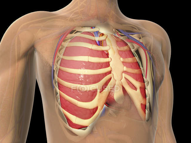
Organs - Stock Photos, Royalty Free Images | Focused from st.focusedcollection.com The final two pairs of ribs are floating ribs and the cartilage of these ribs tends to end ibrahim, af and darwish: The number is the same in both males and females. As such, the 2nd rib's position on the angle of lewy is actually on the same transverse plane as the t4 vertebrae. It is related to the costal pleura, which separates it from the ribs, their costal cartilages, and the innermost intercostal muscles. These anatomical specializations have earned birds their own class in the vertebrate phylum. This page is about anatomy of ribs lungs and diaphragm,contains normal anatomy and flow during the complete examination: These are hooked extensions of bone which help to strengthen the the arrangement of the air sacs, and lungs in birds. The lungs are the primary organs of the respiratory system in humans and many other animals including a few fish and some snails.
Each lung is located near different organs in the body.
Let's take a look at some anatomy of the lungs. Extracardiac anatomy,basics of pediatric anesthesia,the diaphragm,endometriosis & the heart and more. Normal anatomy of the thorax on labeled chest ct: The lungs are the primary organs of the respiratory system in humans and many other animals including a few fish and some snails. Birds have uncinate processes on the ribs. The costal surface of the lung this surface is large, smooth, and convex. During inspiration, the chest and lungs expand in all three planes lungs in infants. Occupying most of the space within the thoracic cavity, the lungs extend laterally from the heart to the ribs on both sides of the chest the bronchioles further branch off into many tiny terminal bronchioles. The anterior border of the lung is formed by the convergence of the mediastinal. The heart and lungs work together to provide oxygen to the cells of the body. The thoracic cage surrounds and protects the heart and lungs in the thoracic cavity. Includes images, video, and free quiz. As such, the 2nd rib's position on the angle of lewy is actually on the same transverse plane as the t4 vertebrae.
The mediastinal surface lies against the mediastinum anteriorly. Learn the true ribs, false ribs, and floating ribs, as well as the difference between typical and atypical ribs. It is related to the costal pleura, which separates it from the ribs and innermost intercostal muscles. Normal anatomy of the thorax on labeled chest ct: The lungs are the essential organs of respiration;
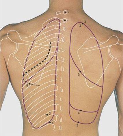
Thorax - Surface Anatomy, 4 Edition from doctorlib.info Neurons in this brain region send signals to the diaphragm and the muscles between the ribs to regulate the contractions which initiate the breathing. The ribs help protect vital organs in the thorax such as the heart and lungs, and they assist with breathing. The infant has several developmental differences in the structure and function of the lung. The lungs are the functional units of respiration and are key to survival. This begins at a somewhat higher level posteriorly, between the third and fifth ribs and runs downward and forward to end in the region of the sixth or seventh costochondral junction. Anatomy of lungs lungs are a pair of respiratory organs situated in thoracic cavity. While these anatomical variations are common and often go unnoticed in otherwise healthy individuals, it's important to distinguish them when reading. They are an important part of the respiratory system and waste management for the there are around 480 million alveoli in the human lungs, according to the department of anatomy of the university of göttingen.
This begins at a somewhat higher level posteriorly, between the third and fifth ribs and runs downward and forward to end in the region of the sixth or seventh costochondral junction.
The pleura is a double‐layered membrane consisting of an inner pulmonary (visceral) pleura, which surrounds each lung, and an outer parietal pleura. Terminal bronchioles are the smallest air tubes in the lungs and terminate at the. Blunt lies above the level of anterior end of 1st rib. The lungs are extending anterior and laterally from the heart to the ribs and posterior to the thoracic spine. Radiological anatomy of the lungs, mediastinal lymph nodes, trachea, bronchi, pleural cavity, heart and pulmonary vessels. Let's take a look at some anatomy of the lungs. Extracardiac anatomy,basics of pediatric anesthesia,the diaphragm,endometriosis & the heart and more. Lungs are a pair of respiratory organs situated in a thoracic cavity. The lungs lie either side of the mediastinum, within the thoracic cavity. It is related to the costal pleura, which separates it from the ribs, their costal cartilages, and the innermost intercostal muscles. The infant has several developmental differences in the structure and function of the lung. The anatomy of bird's respiratory system. Function of lungs and lung anatomy and lung lobes.
The lungs are extending anterior and laterally from the heart to the ribs and posterior to the thoracic spine anatomy of ribs. Vestibular anatomy and neurophysiology online course:


0 Komentar