Upper Leg Tendon Anatomy / Jul 01, 2021 · knee, leg, and foot (overview) how many times have a layman's language and anatomy ever matched?. They originate at the ilium (upper part of the pelvis, or hipbone) and femur (thighbone), come together in a The biceps is attached to the arm bones by. In a complete or serious rupture the tendon of plantaris or another vestigial muscle is harvested and wrapped around the achilles tendon, increasing the strength of the repaired tendon. The biceps is a muscle on the front part of the upper arm. Quadriceps femoris muscle, large fleshy muscle group covering the front and sides of the thigh.
The middle part gives insertion to the achilles tendon The tendon continues its way through the foot by extending over its dorsal surface and finally inserting on the superior surface of the base of the distal phalanx of the hallux. Jul 01, 2021 · knee, leg, and foot (overview) how many times have a layman's language and anatomy ever matched? Rectus femoris, vastus lateralis, vastus medialis, and vastus intermedius. The biceps is a muscle on the front part of the upper arm.
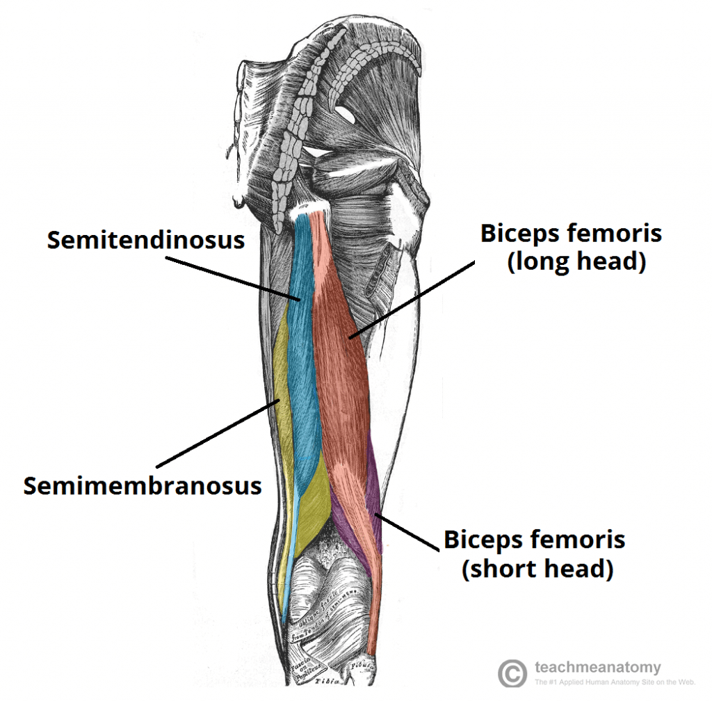
Muscles of the Posterior Thigh - Hamstrings - Damage ... from teachmeanatomy.info Jun 18, 2018 · the upper leg is often called the thigh. The posterior upper leg muscles provide your knees with mobility (extension, flexion and rotation) and strength.they work closely with your quadriceps muscles at the front of your thigh, your gluteal muscles, and your calf muscles to ensure proper movement of your leg and hip. In human anatomy, the peroneus longus (also known as fibularis longus) is a superficial muscle in the lateral compartment of the leg, and acts to evert and plantarflex the ankle. It's the area that runs from the hip to the knee in each leg. They originate at the ilium (upper part of the pelvis, or hipbone) and femur (thighbone), come together in a Quadriceps femoris muscle, large fleshy muscle group covering the front and sides of the thigh. The biceps is a muscle on the front part of the upper arm. The middle part gives insertion to the achilles tendon
The tendon continues its way through the foot by extending over its dorsal surface and finally inserting on the superior surface of the base of the distal phalanx of the hallux.
Jun 18, 2018 · the upper leg is often called the thigh. It's the area that runs from the hip to the knee in each leg. The tendon continues its way through the foot by extending over its dorsal surface and finally inserting on the superior surface of the base of the distal phalanx of the hallux. The biceps is attached to the arm bones by. The upper area is smooth, slopes anteriorly, and supports a bursa, which lies between it and the achilles tendon. The biceps includes a "short head" and a "long head" that work as a single muscle. The purpose of this study was to characterize online video use and understand the role videos play in the learning process of orthopedic residents and practicing surgeons. Jul 01, 2021 · knee, leg, and foot (overview) how many times have a layman's language and anatomy ever matched? Rectus femoris, vastus lateralis, vastus medialis, and vastus intermedius. The biceps is a muscle on the front part of the upper arm. The posterior upper leg muscles provide your knees with mobility (extension, flexion and rotation) and strength.they work closely with your quadriceps muscles at the front of your thigh, your gluteal muscles, and your calf muscles to ensure proper movement of your leg and hip. In a complete or serious rupture the tendon of plantaris or another vestigial muscle is harvested and wrapped around the achilles tendon, increasing the strength of the repaired tendon. The middle part gives insertion to the achilles tendon
They originate at the ilium (upper part of the pelvis, or hipbone) and femur (thighbone), come together in a The middle part gives insertion to the achilles tendon The tendon continues its way through the foot by extending over its dorsal surface and finally inserting on the superior surface of the base of the distal phalanx of the hallux. During an open surgery, an incision is made in the back of the leg and the achilles tendon is stitched together. The upper area is smooth, slopes anteriorly, and supports a bursa, which lies between it and the achilles tendon.
IT Band Injury | Sports Podiatry from www.sportspodiatry.co.uk In a complete or serious rupture the tendon of plantaris or another vestigial muscle is harvested and wrapped around the achilles tendon, increasing the strength of the repaired tendon. During an open surgery, an incision is made in the back of the leg and the achilles tendon is stitched together. Also called the thigh bone, this is the longest bone in the body.it. Quadriceps femoris muscle, large fleshy muscle group covering the front and sides of the thigh. In human anatomy, the peroneus longus (also known as fibularis longus) is a superficial muscle in the lateral compartment of the leg, and acts to evert and plantarflex the ankle. The biceps includes a "short head" and a "long head" that work as a single muscle. The upper area is smooth, slopes anteriorly, and supports a bursa, which lies between it and the achilles tendon. Jun 18, 2018 · the upper leg is often called the thigh.
The middle part gives insertion to the achilles tendon
The tendon continues its way through the foot by extending over its dorsal surface and finally inserting on the superior surface of the base of the distal phalanx of the hallux. The biceps includes a "short head" and a "long head" that work as a single muscle. Rectus femoris, vastus lateralis, vastus medialis, and vastus intermedius. During an open surgery, an incision is made in the back of the leg and the achilles tendon is stitched together. Also called the thigh bone, this is the longest bone in the body.it. The posterior upper leg muscles provide your knees with mobility (extension, flexion and rotation) and strength.they work closely with your quadriceps muscles at the front of your thigh, your gluteal muscles, and your calf muscles to ensure proper movement of your leg and hip. In a complete or serious rupture the tendon of plantaris or another vestigial muscle is harvested and wrapped around the achilles tendon, increasing the strength of the repaired tendon. Quadriceps femoris muscle, large fleshy muscle group covering the front and sides of the thigh. Jul 01, 2021 · knee, leg, and foot (overview) how many times have a layman's language and anatomy ever matched? The biceps is attached to the arm bones by. The upper area is smooth, slopes anteriorly, and supports a bursa, which lies between it and the achilles tendon. It's the area that runs from the hip to the knee in each leg. The purpose of this study was to characterize online video use and understand the role videos play in the learning process of orthopedic residents and practicing surgeons.
They originate at the ilium (upper part of the pelvis, or hipbone) and femur (thighbone), come together in a Quadriceps femoris muscle, large fleshy muscle group covering the front and sides of the thigh. Jul 01, 2021 · knee, leg, and foot (overview) how many times have a layman's language and anatomy ever matched? In a complete or serious rupture the tendon of plantaris or another vestigial muscle is harvested and wrapped around the achilles tendon, increasing the strength of the repaired tendon. The biceps is attached to the arm bones by.
Semimembranosus: Origin, Insertion, Action & Nerve Supply ... from howtorelief.com The tendon continues its way through the foot by extending over its dorsal surface and finally inserting on the superior surface of the base of the distal phalanx of the hallux. Jun 18, 2018 · the upper leg is often called the thigh. Quadriceps femoris muscle, large fleshy muscle group covering the front and sides of the thigh. During an open surgery, an incision is made in the back of the leg and the achilles tendon is stitched together. In a complete or serious rupture the tendon of plantaris or another vestigial muscle is harvested and wrapped around the achilles tendon, increasing the strength of the repaired tendon. The biceps is attached to the arm bones by. They originate at the ilium (upper part of the pelvis, or hipbone) and femur (thighbone), come together in a The purpose of this study was to characterize online video use and understand the role videos play in the learning process of orthopedic residents and practicing surgeons.
The biceps includes a "short head" and a "long head" that work as a single muscle.
The purpose of this study was to characterize online video use and understand the role videos play in the learning process of orthopedic residents and practicing surgeons. Rectus femoris, vastus lateralis, vastus medialis, and vastus intermedius. The posterior upper leg muscles provide your knees with mobility (extension, flexion and rotation) and strength.they work closely with your quadriceps muscles at the front of your thigh, your gluteal muscles, and your calf muscles to ensure proper movement of your leg and hip. In human anatomy, the peroneus longus (also known as fibularis longus) is a superficial muscle in the lateral compartment of the leg, and acts to evert and plantarflex the ankle. The middle part gives insertion to the achilles tendon The biceps includes a "short head" and a "long head" that work as a single muscle. The biceps is a muscle on the front part of the upper arm. Jul 01, 2021 · knee, leg, and foot (overview) how many times have a layman's language and anatomy ever matched? The muscle, the longest and most superficial of the three peroneus muscles , is attached proximally to the head of the fibula and its 'belly' runs down most of this bone. Also called the thigh bone, this is the longest bone in the body.it. During an open surgery, an incision is made in the back of the leg and the achilles tendon is stitched together. The tendon continues its way through the foot by extending over its dorsal surface and finally inserting on the superior surface of the base of the distal phalanx of the hallux. The biceps is attached to the arm bones by.


0 Komentar